We are Science Accelerated
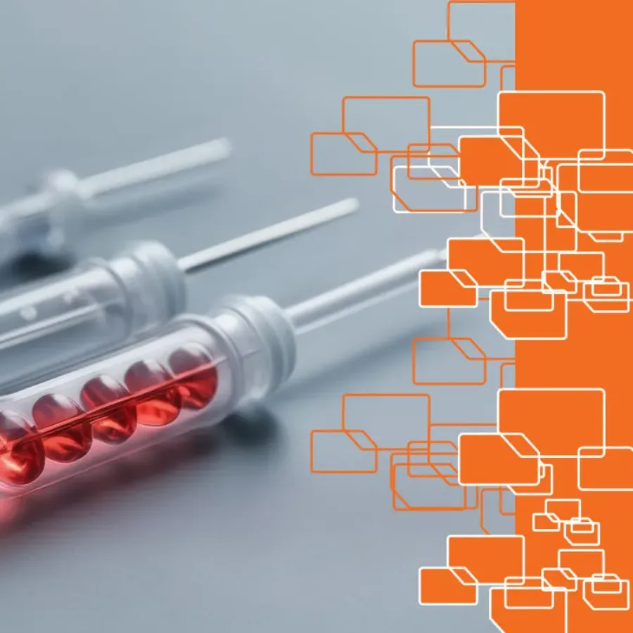
We are KCAS Bio. The full-service bioanalytical CRO solutions partner, helping to accelerate the discovery and development of life-changing drugs smoothly, safely and sustainably.

Bioanalytical CRO service excellence
We take a consultative approach to our discovery and bioanalytical services, giving our clients' confidence their needs are understood, their objectives will be met, and our promises kept. We consistently benchmark, evaluate and optimize our operations so we can help scientific innovators and investors.
At KCAS Bio, we only make promises we can keep.
We combine world-class expertise and innovation to help our clients drive time-critical research forward every day.
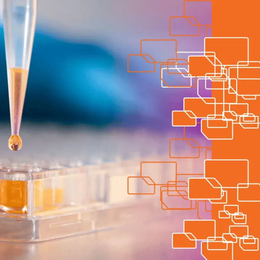
Global biomarker and bioanalytical CRO solutions
KCAS Bio is an established bioanalytical CRO with over 40 years' experience. We are very mindful of the human and financial impact of drug development delays. Through our unique approach to capacity planning, we prepare the right expertise with the right capacity and equipment so that we can keep development moving smoothly. Our clients consider us a reliable partner because we strive to minimize downtime, avoid delays, and keep lead times as short as possible.
Find exactly what you’re looking for
Search for specific services, platforms, instrumentation, or scientific insights.
KCAS Bio scientific insights
 Blogs
Blogs
Dawn Dufield, PhD is the Scientific Officer for Mass Spectrometry and has been with KCAS Bio since 2018. Before joining KCAS Bio, she worked in the quantitative large and small molecule LC-MS/MS field for Pfizer for over 20 years. Dawn has been a pioneer in the Hybrid LCMS field and…
 Blogs
Blogs
Over the past decade, a continued discussion point has been the idea around analyzing samples on Ligand Binding Assay (LBA) platforms in singlet (one well) versus the standard duplicate analysis (Single sample added to two different wells). In the recent M10 Bioanalytical Method Validation Guideline issued for guidance in June…
 Blogs
Blogs
At KCAS Bio, we continuously seek ways to improve our processes with sound, logical, scientific, and business-related methods. One of these areas of evaluation has been sample preparation. We have improved various segments of bioanalysis (drying for unstable analytes, liquid handling, and plate-based assays for increased efficiency). We can also…
Advancing great people and great science
We make nurturing new talent a strategic priority, so that teams are dynamic and constantly improving. We provide a supportive environment for continuous learning and training where you can become your best.
What our clients say
We consistently earn our clients’ trust based on our reputation for transparency, delivering error-free data and keeping our promises.

Tell us how we can help with your project
We've earned our reputation for delivering reliable, error-free data. We understand the importance of speed, flexibility, and consistency and only make promises we can keep.