Oncology CRO Services
KCAS has the expertise to support the Immuno-oncology field. We have expertise in flow cytometry, ELISPOTs, cell based assays and multiple biomarkers for the development of new drugs. Whether it is in-vitro discovery method or support of a phase III trial, we can assist you. We provide reliable and defendable data while continuing to help our partners develop drugs for the advancement of world health.

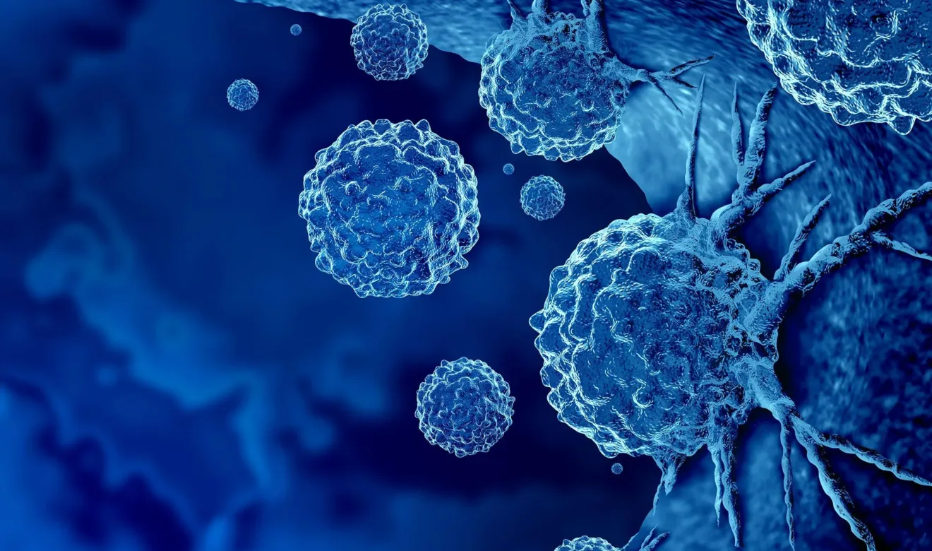
Rapid growth in immuno-oncology
Immuno-oncology is a growing field. The goal is to augment the patient’s immune system to attack the cancer cells. The current lines of treatment for cancer are radiation, chemotherapy, and surgical resection. If these treatments do not work, physicians can seek alternative treatments, such as immune-oncology therapeutics. Two emerging immune-oncology therapeutic modalities are saving lives: antibody drug conjugates (ADCs) and cellular therapies.
Oncology research, by leading scientists
Developing a reliable immune assay requires significant scientific knowledge and optimization time investment. This also requires precise and consistent handling for repeatable results to reduce variations in assay performance and unreliable data. KCAS has the experience and the expertise to offer you the tools you need for a successful program such as flow cytometry
We employ rigorous quality practices that go beyond our competition. Processes are continuously improved to ensure we consistently deliver a complete outsourcing experience to clients.
KCAS Bio provides high quality science and data services that have benefits for the entire drug development journey.
Agile, responsive, and easy to work with
We prepare and adapt our oncology research based on a deep understanding of your drug development ambitions and wider business objectives.
KCAS Bio scientific insights
 Blogs
Blogs
Immuno-oncology is a growing field. The goal is to augment the patient’s immune system to attack the cancer cells. The current lines of treatment for cancer are radiation, chemotherapy, and surgical resection. If these treatments do not work, physicians can seek alternative treatments, such as immune-oncology therapeutics. Two emerging immune-oncology…
 Blogs
Blogs
Spectral flow has come a long way in recent years, transforming from a mere whisper of possibility to an essential tool in bioanalysis. Initially gaining traction in academic institutions, it quickly caught the attention…

Tell us how we can help with your project
We've earned our reputation for delivering reliable, error-free data. We understand the importance of speed, flexibility, and consistency and only make promises we can keep.